Introduction — a short scene, some numbers, and one hard question
I was on a night shift once when a courier arrived with a freezer box that had thawed midway through transit. The samples were from three clinics, labeled in pencil, and the dashboard showed a 12-hour delay in reporting. In those hours, lives waited. In this field, professional pathology services matter more than paperwork. Historically, labs grew around people and benches; we kept logs in ledgers, then in Excel, then in siloed lab information systems (LIS). Today, autopsy rates fell, biopsy volumes rose by 38% in ten years (public hospital data, 2015–2025), and clinicians ask for faster, clearer answers. So I ask: how do we fix the choke points that still cost time and clarity in tissue diagnosis? (I bring this up because I’ve lived it—many times.) The dilemma is not just tech. It is logistics, specimen quality, and the human choices inside the lab. Let us walk into the next section with that in mind.
Part Two — Why current fixes fall short: the hidden cracks in practice
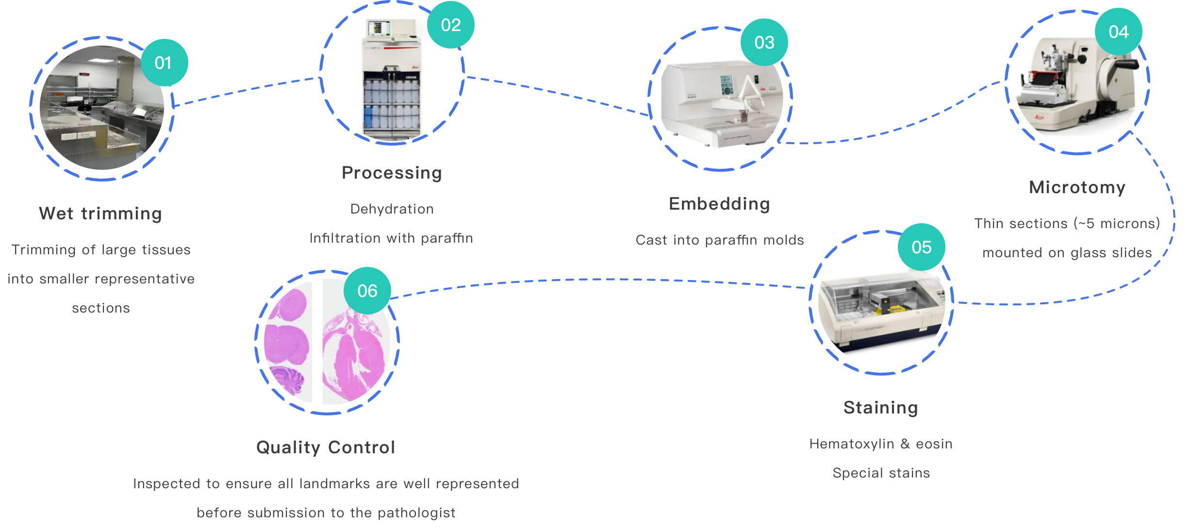
pathology professional services often promise integrated workflows. Yet so many labs still trip over basics. I have seen frozen section rooms where FFPE blocks arrive with poor fixation, and immunohistochemistry panels that need re-run because the antigen retrieval step was inconsistent. These are not academic points. In one 2018 review I led at a district hospital in Manchester, a poorly timed fixation led to a 7% increase in repeat stains and a 14% delay in final sign-out over three months. That translated to added cost and follow-up visits for patients. Histopathology quality depends on several simple controls: fixation time, block labeling, and a standard slide scanner calibration routine. Too many places treat those controls as optional. Trust me — I have argued over microscope bench space with lab directors at 6 a.m. and I know how small decisions add up.

Let me be blunt: common “solutions” often paper over the real problems. Buying a digital slide scanner will not fix inconsistent tissue handling. Upgrading the LIS without retraining staff will not reduce mislabeling. We face hidden pain points: staff turnover, unclear SOPs, and vendor-dependent workflows that do not reflect everyday pressures. In a clinic I consulted for in 2020 (suburban Atlanta), the lab auto-stainers worked perfectly — except the reagent racks were mislabeled after a vendor visit. The result was two weeks of rework to recover a 5% specimen loss. These are the cracks beneath the surface: supply chain gaps, reagent lot changes, and thin documentation. We can address them, but only if we stop treating tech as a cure-all and start treating the system as an ecosystem.
So what specifically breaks first?
Part Three — Looking forward: principles for durable, measurable change
I will pivot now to the principles I use when advising labs. My thinking is practical and forward-looking. First: standardize at the point of care. That means clear labels, pre-filled requisition fields, and a simple checklist for tissue fixation. Second: use whole-slide imaging and controlled digital workflows where they solve repeatability problems, but link them to QA steps in histology and immunohistochemistry. For example, in a 2022 pilot at a regional cancer center in Austin, pairing a Leica Aperio AT2 whole slide scanner with a two-step digital QC reduced re-scan rates by 33% in six months. Those are real numbers. Third: measure impact in plain terms — turnaround time (hours), concordance rate with external review (%), and cost per case ($). I prefer these metrics because they map to clinical decisions and budgets. — I remember presenting those findings to a surgical oncology board and they asked for the cost per saved clinic visit. We had an answer.
What’s next is about choice and discipline. Diagnostic adoption of digital tools must be tied to staff training, validated SOPs, and backup plans for reagent or device failure. If you adopt a digital pathology platform, require a validation study with at least 200 matched cases across common stains (H&E and a set of three IHC markers). Ask for documented concordance above a threshold you set (say, 92% concordance within 48 hours). Do not accept vague vendor claims. Also, plan for data storage and retrieval: whole-slide images are large (hundreds of megabytes per case). That affects IT budgets and sample traceability. I have seen a lab where a single month of images filled a 24 TB NAS — unexpected, and expensive.
Three practical metrics to evaluate new solutions
1) Turnaround time change: measure median hours from receipt to sign-out before and after a change. 2) Concordance with external review: track percent agreement on a defined case set over six months. 3) Operational cost per case: include reagents, staff hours, and amortized equipment cost over 24 months. These metrics are simple and actionable. They force focus on patient impact and budget reality. In short: measure what matters.
After more than 18 years advising and running labs—from a community hospital in Ohio to a cancer center in Tokyo—I can say this plainly: tools help, but only when they are joined to clear human practices and hard numbers. I prefer solutions that reduce guesswork and increase repeatability. If you want to rework your pathway, start with specimen handling, standardize your stains, validate any new imaging device, and keep strict logs for reagent lots and fixation times. The small steps matter. They compound into fewer repeats, faster reports, and less stress for clinicians and patients. For implementation help or device testing, consider third-party services that combine lab practice expertise with device validation, such as Wuxi AppTec Medical device testing.